
Trust the myope: symptoms as the trigger for treatment in mCNV
A 52-year-old man with high myopia presented in 2018 with a scotoma superotemporal to fixation in the left eye. Visual acuity measured 6/6 right and 6/9 left. Fundus examination revealed a retinal haemorrhage, and OCT demonstrated subretinal hyperreflective material (SRHM) at an inferonasal juxtafoveal location. He was diagnosed with myopic choroidal neovascularisation (mCNV) and treated with intravitreal anti-VEGF, with resolution of symptoms and improvement in visual acuity to 6/6.
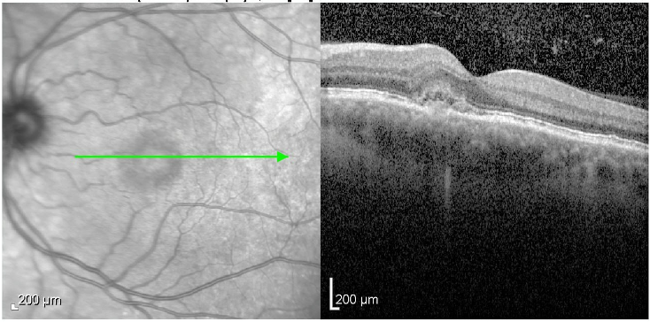
Macular OCT - left eye, 2018. Subretinal hyperreflective material (SRHM) in the inferonasal juxtafoveal region with elevation of the overlying neurosensory retina, consistent with active type 2 myopic CNV.
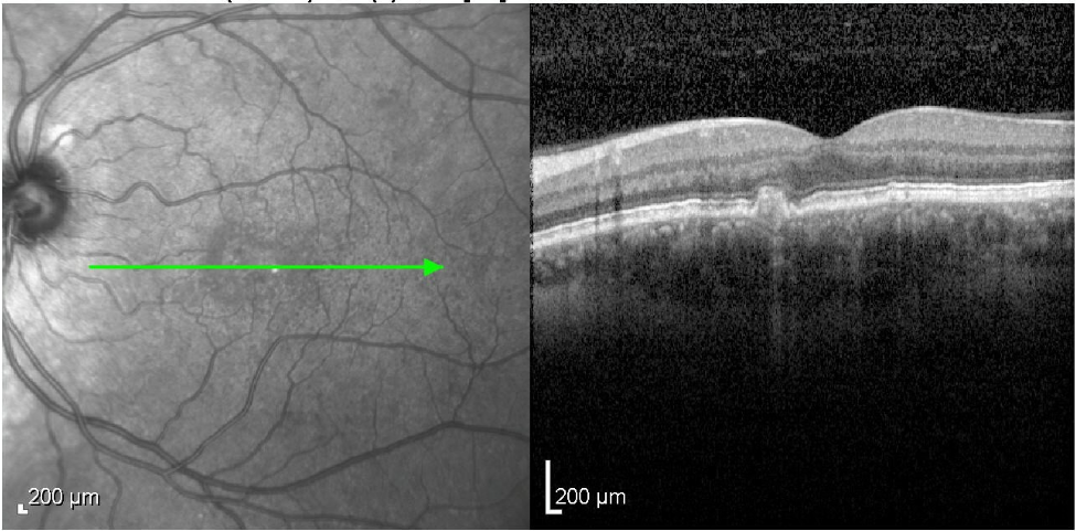
Macular OCT - left eye, post-treatment. Resolution of SRHM with residual fibrosis and a hypertransmission defect beneath the treated lesion. Visual acuity recovered to 6/6.
He remained asymptomatic for eight years before presenting on an SOS basis in September 2026 with a few weeks of recurrent distortion in the same part of his visual field. Visual acuity was maintained at 6/6. Fundus examination showed no haemorrhage. The structural OCT was similar to the post-treatment scan, showing apparent stable fibrosis with underlying hypertransmission, without obvious SRHM or fluid - an appearance that could easily be attributed to quiescent treated disease.
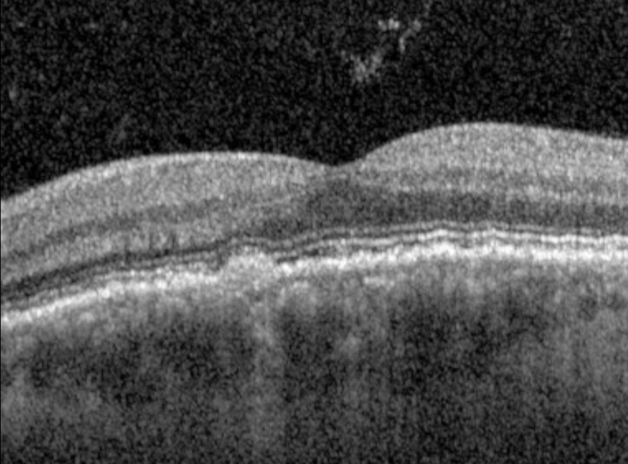
Macular OCT - left eye, September 2026. Apparent stable fibrosis with underlying hypertransmission defect. No haemorrhage, SRHM or subretinal fluid identified on structural OCT alone.
OCT angiography (OCT-A) resolved the diagnostic uncertainty, demonstrating a lacy neovascular network in the avascular complex slab corresponding to the area of fibrosis - the characteristic appearance of active CNV that was invisible on structural OCT alone. Intravitreal anti-VEGF was administered the same day.
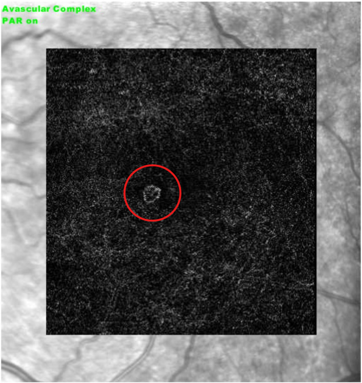
OCT angiography - avascular complex slab, left eye, September 2026. A small but clearly defined lacy ring-like neovascular network (circled) within the avascular complex, confirming active CNV recurrence in the region of apparent stable fibrosis. This signal was invisible on structural OCT alone.
The classic presentation of myopic CNV - haemorrhage and subretinal hyperreflective material on OCT - reflects the predominantly type 2 (sub-retinal, classic) nature of the membrane, which breaks through the RPE and grows into the subretinal space. This is in contrast to type 1 CNV (sub-RPE, occult), which remains beneath the RPE and tends to present more subtly. While the type 1/type 2 distinction carries less direct management weight in myopic CNV than it once did in AMD - where it historically influenced whether PDT was indicated before anti-VEGF superseded it as first-line treatment - understanding why haemorrhage and SRHM predominate helps explain both the classic presentation and why their absence at recurrence can be misleading. The OCT-A here shows a lacy, frond-like neovascular network consistent with type 2 recurrence, but at an early stage before sufficient exudate has accumulated to be visible as SRHM on structural OCT. This is an important lesson: OCT-A can detect active type 2 CNV recurrence before it declares itself on B-scan imaging. Both types respond well to anti-VEGF, as demonstrated by Huang et al. (Retina 2025) , with equivalent recurrence rates and injection numbers regardless of CNV type.
This case illustrates two important points. First, as EyeWiki notes and expert consensus supports, a history of metamorphopsia in a myopic patient should arouse suspicion of CNV - and in a patient with prior treated mCNV presenting with recurrent distortion in precisely the same location, the clinical case for treatment is strong even before imaging is reviewed. There are no dedicated society guidelines for mCNV, but the key trials - RADIANCE and MYRROR - established anti-VEGF as first-line treatment. Expert sources including the AAO and a EURETINA systematic review explicitly include metamorphopsia and visual symptoms alongside structural imaging findings as criteria for treatment - supporting the principle that in mCNV, the history is as important as the scan. In this case, I would have been inclined to treat on clinical suspicion alone; the OCT-A simply confirmed what the history had already suggested. Second, OCT-A is a valuable adjunct in the surveillance of treated myopic CNV, capable of detecting recurrent neovascular activity before it declares itself on structural OCT.