
Cotton wool spot or something else? An inner retinal lesion in context
A 65-year-old man with a known history of tuberous sclerosis complex (TSC) was referred by his optometrist following an incidental OCT finding at a routine sight test. He had no visual symptoms, Amsler distortions, or visual field loss. Visual acuity corrected to 6/7.5 bilaterally. His medical history was significant: he had undergone total nephrectomy in 1986 for a benign renal growth, developed end-stage chronic kidney disease, and had recently received a renal transplant in November 2025, for which he was on immunosuppression including mycophenolate mofetil, tacrolimus, and prednisolone. He also had type 2 diabetes and hypertension.
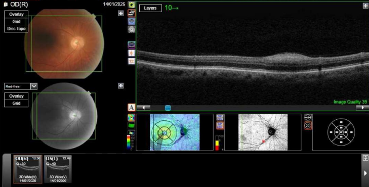
Macular OCT - right eye, submitted with the original referral. A striking hyperreflective thickening of the inner retina temporal to the disc, with no corresponding visible lesion on the adjacent fundus photograph - the finding that prompted referral.
The optometrist described a large area of bright thickening in the inner retina temporal to the macula on OCT, with no visible lesion, scar, or haemorrhage on fundoscopy or fundus photography. The referral initially had to be returned to the GP for resubmission with the OCT images attached - the images had not been included in the original referral - resulting in a short delay before the patient could be accepted and booked. He was seen in a medical retina clinic within eight weeks of the completed referral.
Tuberous sclerosis complex is an autosomal dominant multisystem disorder caused by mutations in the TSC1 or TSC2 tumour suppressor genes, resulting in benign hamartomatous growths in multiple organs including the brain, kidneys, skin, lungs, and eyes. It is a phakomatosis - one of a group of neurocutaneous syndromes with characteristic lesions in the nervous system and skin. This patient’s renal history - nephrectomy in 1986 for a benign growth and subsequent end-stage renal failure requiring transplantation - is highly consistent with TSC-related renal angiomyolipomas, a recognised and potentially serious extraocular manifestation of the condition.
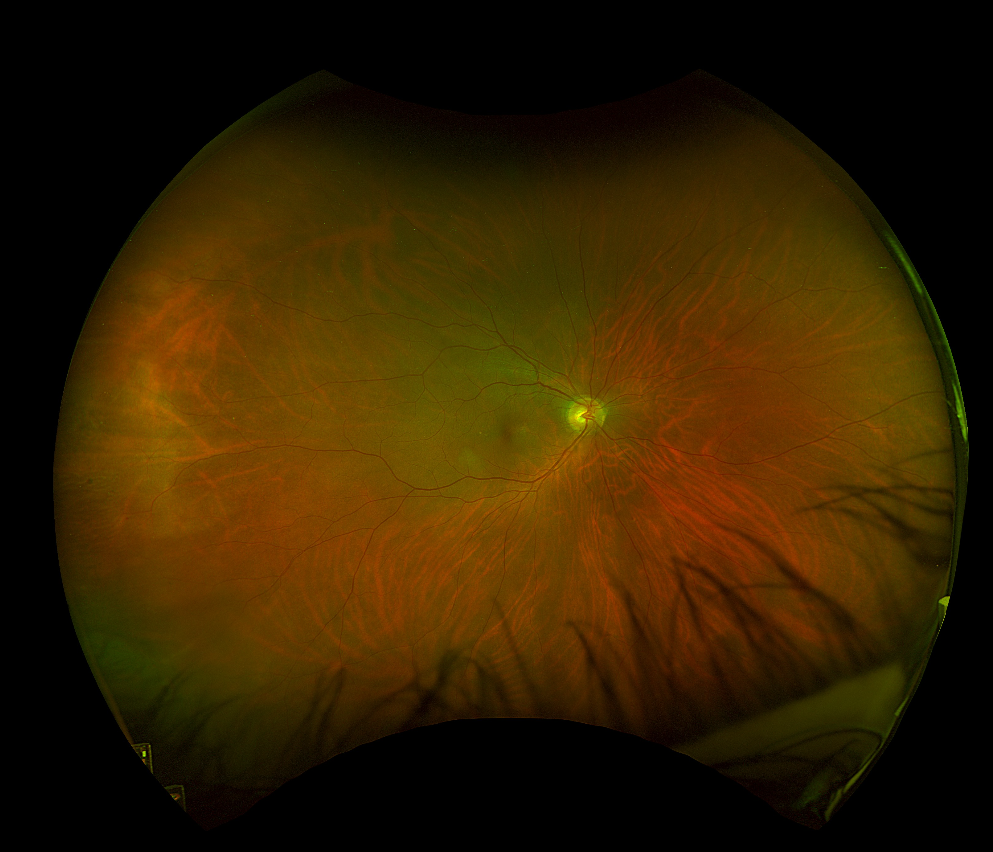
Widefield colour fundus photograph - right eye. A slightly raised, pale lesion is visible temporal to the optic disc, subtle on colour photography but conspicuous on OCT.
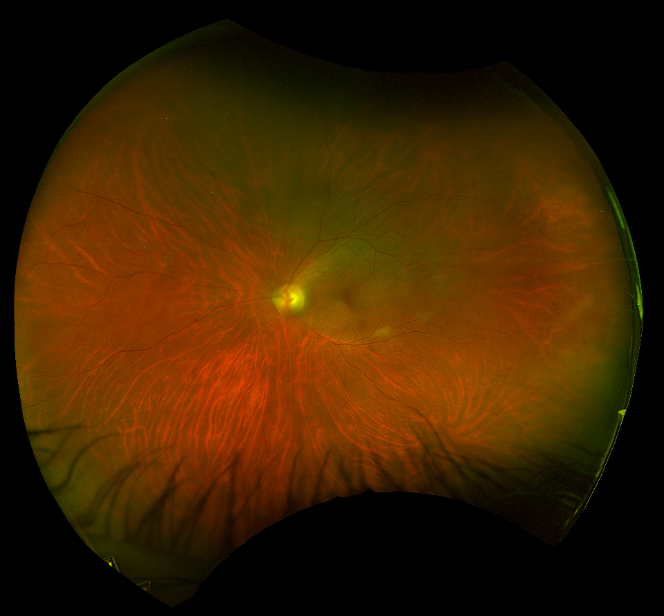
Widefield colour fundus photograph - left eye. A corresponding lesion near the optic disc confirming bilateral involvement.
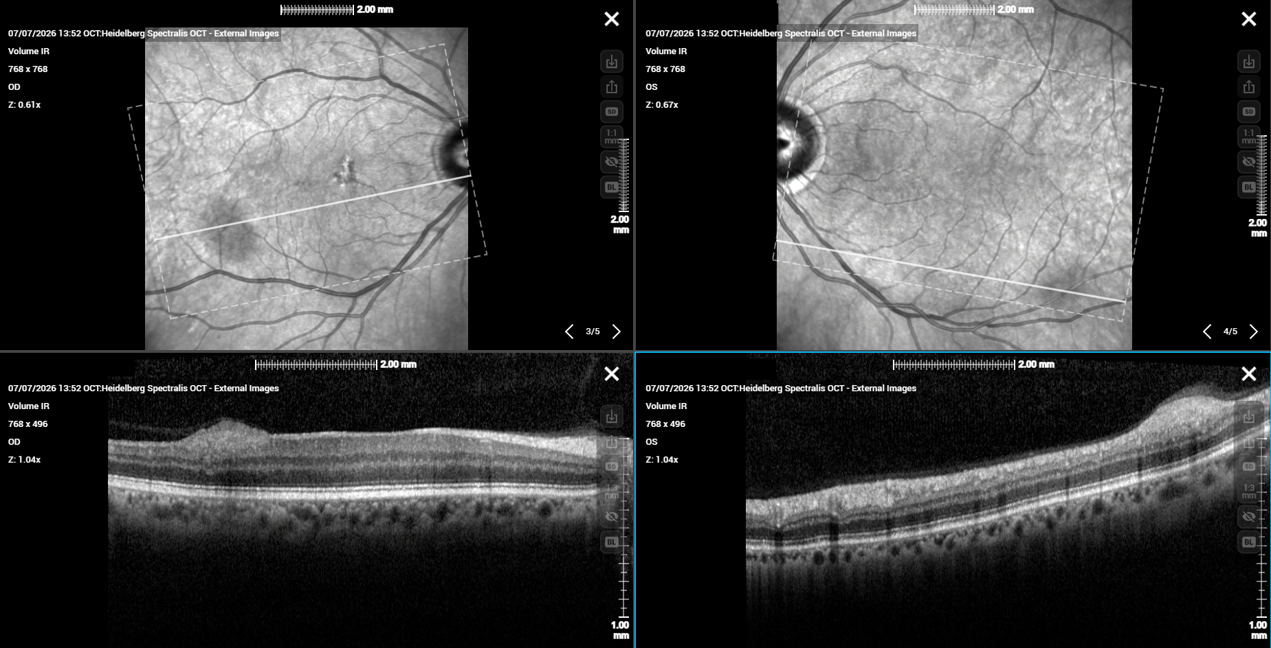
Bilateral macular OCT - right (left panel) and left (right panel) eyes. Right eye: hyperreflective lesion projecting from the nerve fibre layer and inner limiting membrane surface. Left eye: corresponding hyperreflective lesion confirming bilateral involvement.
The OCT demonstrates the characteristic appearance of retinal astrocytic hamartomas (RAH) - hyperreflective lesions arising in the nerve fibre layer and projecting from the inner limiting membrane surface. Shields et al. (Ophthalmology 2016) described two OCT patterns: solid hyperreflective thickening projecting from the nerve fibre layer and inner limiting membrane surface, as seen here, and a moth-eaten appearance with optically empty spaces, which tends to occur in larger or more calcified lesions. This surface-projecting profile distinguishes RAH from cotton wool spots, which produce a more diffuse hyperreflective thickening spreading through multiple inner retinal layers - a distinction illustrated in the hypertensive retinopathy case on this site. The differential diagnosis of a bright inner retinal lesion on OCT in this patient warrants careful consideration. Cotton wool spots (CWS) - areas of focal nerve fibre layer infarction - produce a similar hyperreflective inner retinal thickening on OCT and would be entirely plausible given his diabetes, hypertension, and chronic renal disease. However, several features argue firmly against CWS here: the lesions are present bilaterally in a stable configuration, are visible on colour photography as slightly raised pale lesions (CWS appear as fluffy white superficial patches that fade within weeks to months), the patient had no visual symptoms, and critically, his known history of tuberous sclerosis provides the unifying diagnosis. CWS also typically occur in the setting of acute systemic decompensation or active vascular disease, whereas these lesions had been stable on the optometrist’s own serial retinal photographs, with the April 2025 colour images showing the same appearance - OCT simply had not been performed at that visit. RAH are benign glial tumours occurring in approximately 40-50% of patients with TSC, typically bilateral and multiple, most commonly located in the posterior pole.
Visual acuity was 6/6 bilaterally at clinic review. The lesions were documented with retinal imaging and the patient was reassured and discharged. No treatment was indicated. This case illustrates two points: first, that OCT can detect RAH before they are visible on conventional fundoscopy, making it a valuable tool in patients with known TSC; and second, that referrals accompanied by images allow prompt and accurate triage - the initial delay in this case, caused by images being omitted from the referral, is a reminder that imaging should always be submitted alongside the clinical letter.